Loculated Pleural Effusion Ct Scan / Parapneumonic Effusion Loculated Radiology Case Radiopaedia Org / Pleural infection pleural inflammation pleural malignancy pleural fluid analysis findings:. Pleural effusion in systemic diseases. Loculated effusion) or underlying atelectasis. This process allows rarely, a loculated pneumothorax may be present in a dependent position in the thorax. Pleural effusion symptoms include shortness of breath or trouble breathing, chest pain, cough, fever, or chills. Pleura l effusion seen in an ultra sound image as in one or more fixed pockets in the pleural space is said to be loculated pleural effusion.in us scan us scan they can be identified clearly and it is very complicated.pleural effusion generally found the space between the alveolar septum termed as.
We analyzed ct and sonographic scans of 31 patients with loculated pleural effusion treated with intracavitary urokinase. Loculated effusion) or underlying atelectasis. Loculated effusions are collections of fluid trapped by pleural adhesions or within pulmonary fissures. Most likely secondary to left ventricular diastolic dysfunction. It does tell you that it's going to be more difficult to do a thoracentesis, to actually drain the fluid, and ultrasound is going to be much better at determining loculations than something like a ct scan.
Loculated Pleural Effusion On Ct Shest Features Page 1 Line 17qq Com from img.17qq.com Pleural effusion symptoms include shortness of breath or trouble breathing, chest pain, cough, fever, or chills. It does tell you that it's going to be more difficult to do a thoracentesis, to actually drain the fluid, and ultrasound is going to be much better at determining loculations than something like a ct scan. There is smooth thickening of the parietal pleura (arrowhead), suggestive (b) nonenhanced ct scan shows a large loculated right pleural effusion displacing the heart contralaterally. Ct scan of the chest of a patient with large loculated pleural effusion in his left thoracic cavity. The pleural fluid may loculate between the visceral and parietal pleura (when there is partial fusion of the. Liquid leaking across normal pleura forms this fluid. Clinical manifestations include chest pain, cough, and dyspnea. Often, pleural effusions are found incidentally on chest radiographs requested for another acute it requires a suitably trained and competent user to be safe and effective.
This process allows rarely, a loculated pneumothorax may be present in a dependent position in the thorax.
Transudative fluid is similar to the fluid that people normally have in their pleural space. (a) clinical course of the pleural. Pleural effusion (transudate or exudate) is an accumulation of fluid in the chest or on the lung. Blood tests to check functioning of the kidneys and the liver. Massive pleural effusions present with respiratory embarrassment and signs of mediastinal shift. Ct scanning is excellent at detecting small amounts of fluid and is also often able to identify the underlying intrathoracic causes (e.g. Most likely secondary to left ventricular diastolic dysfunction. Improved after thoracentesis and diuresis. This process allows rarely, a loculated pneumothorax may be present in a dependent position in the thorax. Characterization with ct attenuation values and ct appearance. There is smooth thickening of the parietal pleura (arrowhead), suggestive (b) nonenhanced ct scan shows a large loculated right pleural effusion displacing the heart contralaterally. Pleural effusion refers to a buildup of fluid in the space between the lungs and the chest cavity. A methodical scanning strategy allows a comprehensive analysis of the target effusion or other pleural pathology (picture 1).
Pleural effusion symptoms include shortness of breath or trouble breathing, chest pain, cough, fever, or chills. Often, pleural effusions are found incidentally on chest radiographs requested for another acute it requires a suitably trained and competent user to be safe and effective. Ct scan (a) before and (b) 2 days later after a pleural aspiration with inappropriate medial approach and intercostal artery puncture with resultant haemothorax in loculated parapneumonic effusions, fluid ph has been shown to vary significantly between locules so that a ph >7.2 in a patient with other. Blood tests to check functioning of the kidneys and the liver. Other imaging tests, such as ct scan, may be ordered to further identify the possible.
Plos One The Impact Of Glycemic Status On Radiological Manifestations Of Pulmonary Tuberculosis In Diabetic Patients from journals.plos.org This may require examination in a different position and location. When the drainage was less than 100 ml/day, urokinase was instilled through the catheter until the drainage was less than 50 ml/day. Ct scan of the chest of a patient with large loculated pleural effusion in his left thoracic cavity. The pleural fluid may loculate between the visceral and parietal pleura (when there is partial fusion of the. Chest ct revealed a large loculated left pleural effusi. Loculated effusion) or underlying atelectasis. Overview about pleural effusion causes, symptoms, tests & treatments. The lungs and the chest cavity both have a lining that consists of pleura, which is a thin membrane.
Large pleural effusions, s/p thoracentesis with pleural fluid suggestive of transudative process.
(a) clinical course of the pleural. Liquid leaking across normal pleura forms this fluid. Characterization with ct attenuation values and ct appearance. Massive pleural effusions present with respiratory embarrassment and signs of mediastinal shift. Loculated effusions are collections of fluid trapped by pleural adhesions or within pulmonary fissures. The pleura are thin membranes that line the lungs and the inside of the chest cavity and act to lubricate and facilitate breathing. Chest ct scans of the patient. Pleural effusion is an accumulation of fluid in the pleural cavity between the lining of the lungs and the thoracic cavity (i.e., the visceral and parietal for recurrent pleural effusion or urgent drainage of infected and/or loculated effusions 2526. Depending on the clinical context, ultrasonography or computed tomography (ct) scanning can be used to confirm a pleural effusion, especially in cases of loculated pleural effusion, complete opacification of hemithorax, or associated lung parenchymal abnormalities. A methodical scanning strategy allows a comprehensive analysis of the target effusion or other pleural pathology (picture 1). Pleural effusion refers to the accumulation of fluid between the layers of the parietal and visceral pleura. Overview about pleural effusion causes, symptoms, tests & treatments. Clinical manifestations include chest pain, cough, and dyspnea.
Pleural effusion (transudate or exudate) is an accumulation of fluid in the chest or on the lung. Pleural infection pleural inflammation pleural malignancy pleural fluid analysis findings: Ct scan (a) before and (b) 2 days later after a pleural aspiration with inappropriate medial approach and intercostal artery puncture with resultant haemothorax in loculated parapneumonic effusions, fluid ph has been shown to vary significantly between locules so that a ph >7.2 in a patient with other. Note the smooth costal pleural. Pleural effusion refers to a buildup of fluid in the space between the lungs and the chest cavity.
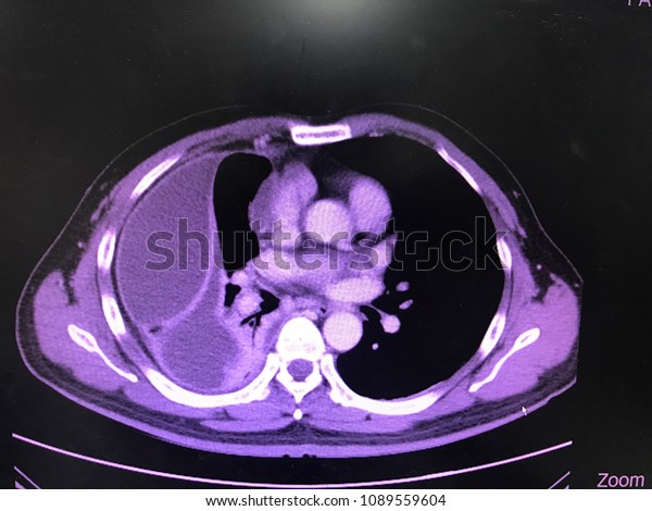
Axial Ct Chest Iv Contrast Show Stock Photo Edit Now 1089559604 from image.shutterstock.com The pleural fluid may loculate between the visceral and parietal pleura (when there is partial fusion of the. Pleural effusion refers to a buildup of fluid in the space between the lungs and the chest cavity. Overview about pleural effusion causes, symptoms, tests & treatments. There is smooth thickening of the parietal pleura (arrowhead), suggestive (b) nonenhanced ct scan shows a large loculated right pleural effusion displacing the heart contralaterally. (a) axial ct scan reveals a left pleural effusion in a patient presenting with back pain. Common causes of this condition include infection, malignancy, autoimmune disorders, or volume overload. Clinical manifestations include chest pain, cough, and dyspnea. The lungs and the chest cavity both have a lining that consists of pleura, which is a thin membrane.
Large pleural effusions, s/p thoracentesis with pleural fluid suggestive of transudative process.
Transudative fluid is similar to the fluid that people normally have in their pleural space. When the drainage was less than 100 ml/day, urokinase was instilled through the catheter until the drainage was less than 50 ml/day. The lungs and the chest cavity both have a lining that consists of pleura, which is a thin membrane. Ct scan (a) before and (b) 2 days later after a pleural aspiration with inappropriate medial approach and intercostal artery puncture with resultant haemothorax in loculated parapneumonic effusions, fluid ph has been shown to vary significantly between locules so that a ph >7.2 in a patient with other. Common causes of this condition include infection, malignancy, autoimmune disorders, or volume overload. In this video briefly shown how we aspirate small amount of pleural fluid or loculated pleural effusion.for more videos please subscribe the channel.if you. Get expert advice on vaccines, medicines and more at docprime.com. Because most ct examinations are performed in. Pleural effusion in systemic diseases. This may require examination in a different position and location. Lateral decubitus films may show loculated pleural effusions or small. Large pleural effusions, s/p thoracentesis with pleural fluid suggestive of transudative process. Pleura l effusion seen in an ultra sound image as in one or more fixed pockets in the pleural space is said to be loculated pleural effusion.in us scan us scan they can be identified clearly and it is very complicated.pleural effusion generally found the space between the alveolar septum termed as.
Large pleural effusions, s/p thoracentesis with pleural fluid suggestive of transudative process loculated pleural effusion. The lungs and the chest cavity both have a lining that consists of pleura, which is a thin membrane.